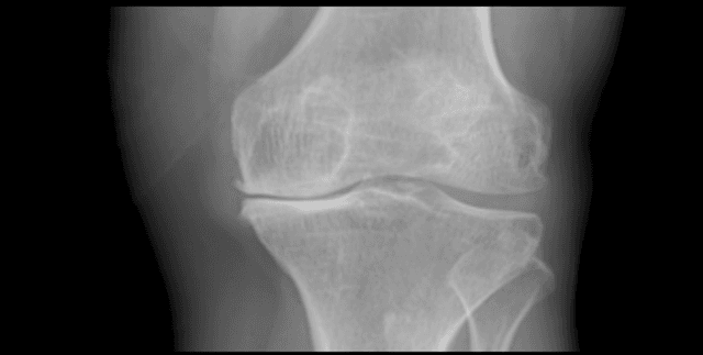
Digital Knee X-ray Images
Healthcare Providers & Services Utilization
Tags and Keywords
Trusted By


"No reviews yet"
Free
About
This curated dataset features 1,650 high-quality digital X-ray images of knee joints, meticulously sourced from leading hospitals and diagnostic centres. The collection is designed to accelerate the development of automated diagnostic tools for knee osteoarthritis (OA), a condition impacting millions globally where accurate diagnosis is crucial for successful treatment. Each image is expertly labeled using the Kellgren and Lawrence (K&L) grading system, recognised as the gold standard for assessing OA severity through radiographic features. This robust, labeled collection allows researchers to merge medicine and technology for improved health outcomes.
Columns
The associated metadata file, Digital Knee X-ray Images.csv, includes details structured across three key columns, in addition to the primary K&L grade annotations included separately:
- MedicalExpert-I: Used to identify the first medical expert involved in the annotation process. This column displays 2 unique values.
- Subdirectory: Provides information on file organisation and categorisation, with '0Normal' being the most frequently occurring value at 20%. It contains 5 unique values.
- File Count: Numerical information relating to the total number of values, with a mean of 330, and values ranging from a minimum of 206 up to a maximum of 514.
Distribution
The dataset consists of 1,650 digital knee X-ray image files. These images are captured in grayscale and have an 8-bit depth, having been processed using a PROTEC PRS 500E X-ray machine. The data is organised within a main directory containing all 1,650 image files. Expert annotations, specifically the K&L grades labeled by two medical professionals, are provided either embedded within the filenames or within a separate metadata file, such as a CSV or text file. The accompanying CSV metadata file is 330 B in size.
Usage
This collection is perfectly suited for several high-impact applications, including:
- Developing models for the automated Kellgren and Lawrence grading of knee osteoarthritis.
- Creating computer-aided diagnosis (CAD) tools specifically designed to assist clinicians.
- Designing advanced pattern recognition algorithms focused on spotting OA-specific features.
- Advancing medical image processing techniques for X-ray analysis and enhancement.
- Innovating techniques using cartilage region of interest (ROI) extraction to derive deeper OA insights based on pixel density.
Coverage
The data focuses exclusively on digital X-ray images of the knee joint. The data was sourced from top hospitals and diagnostic centres. This work was brought forth by Rani Channamma University, located in India.
License
Creative Commons Attribution 4.0 International (CC BY 4.0)
Who Can Use It
This product is highly valuable for:
- Data scientists and machine learning engineers: For training advanced pattern recognition and image classification models.
- Researchers and innovators: Seeking labeled medical data to advance medical image analysis and automated detection systems.
- Medical professionals: Utilizing the resulting Computer-Aided Diagnosis (CAD) tools in clinical settings for rapid assessment.
Dataset Name Suggestions
- Digital Knee X-ray Images
- Annotated Knee OA Grading Dataset
- K&L Graded Knee Radiography Collection
- Automated OA Detection Dataset
Attributes
Original Data Source: Digital Knee X-ray Images
Loading...
