Dark Mode

MRI Brain Tumour Classification Data
Patient Health Records & Digital Health
Tags and Keywords
Trusted By


"No reviews yet"
Free
About
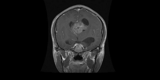
Brain magnetic resonance imaging (MRI) data curated specifically for image classification tasks in health and cancer research. The data consists of MRI images categorised into four distinct groups: Glioma, meningioma, pituitary, and 'no tumor' cases. The primary function of this collection is to facilitate the training and evaluation of machine learning models aimed at identifying different brain tumour types. The data is pre-divided into Training and Testing subsets to support immediate classification work.
Columns
The metadata structure relating to the image breakdown includes four main fields:
- Dataset: Specifies whether the images belong to the Training or Testing set.
- Subdirectory: Denotes the specific tumor type or category (e.g., glioma, meningioma, notumor, pituitary).
- File Count: Indicates the number of images available within that specific subdirectory/dataset split.
- Description: A field for accompanying notes, though often null or unavailable in the structural breakdown.
Distribution
The image data is structured for classification with a significant split between Training and Testing sets, with Testing accounting for 56% of the overall structure and Training accounting for 44%.
The file counts across the subdirectories are detailed as follows:
Training Set:
- glioma: 1,321 images
- meningioma: 1,339 images
- notumor: 1,595 images
Testing Set:
- glioma: 300 images
- meningioma: 306 images
- notumor: 405 images
- pituitary: 300 images
The minimum number of images in any single subdirectory split is 300, and the maximum is 1,595.
Usage
This collection is ideal for developing and benchmarking deep learning models for medical image classification. Specific uses include:
- Training neural networks to differentiate between various brain tumour types (Glioma, Meningioma, Pituitary).
- Creating diagnostic support tools for medical practitioners.
- Research into the severity and characteristics of different brain pathologies using MRI scans.
- Educational purposes in data science and biomedical engineering curricula.
Coverage
The data covers MRI images of brains focusing on four distinct diagnostic categories: Glioma, meningioma, pituitary tumors, and healthy (no tumor) subjects. The expected update frequency for this specific version of the dataset is stated as 'Never'. There is no explicit geographical, temporal, or demographic information provided within the scope of the sources.
License
Attribution 4.0 International (CC BY 4.0)
Who Can Use It
- Data Scientists/Machine Learning Engineers: Utilising supervised learning techniques for image classification model development.
- Medical Researchers: Investigating pattern recognition in pathological MRI images.
- Health Technology Developers: Building prototypes for automated diagnostic assistance.
Dataset Name Suggestions
- Brain MRI Tumour Image Classification Data
- Multi-Class Brain Tumor MRI Dataset
- Glioma, Meningioma, and Pituitary MRI Data
- Medical Deep Learning Image Set
Attributes
Original Data Source:MRI Brain Tumour Classification Data
Loading...